Strain Pattern Ecg
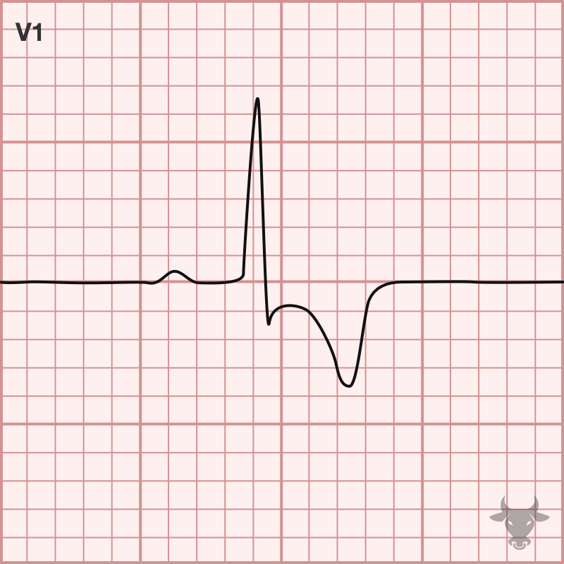
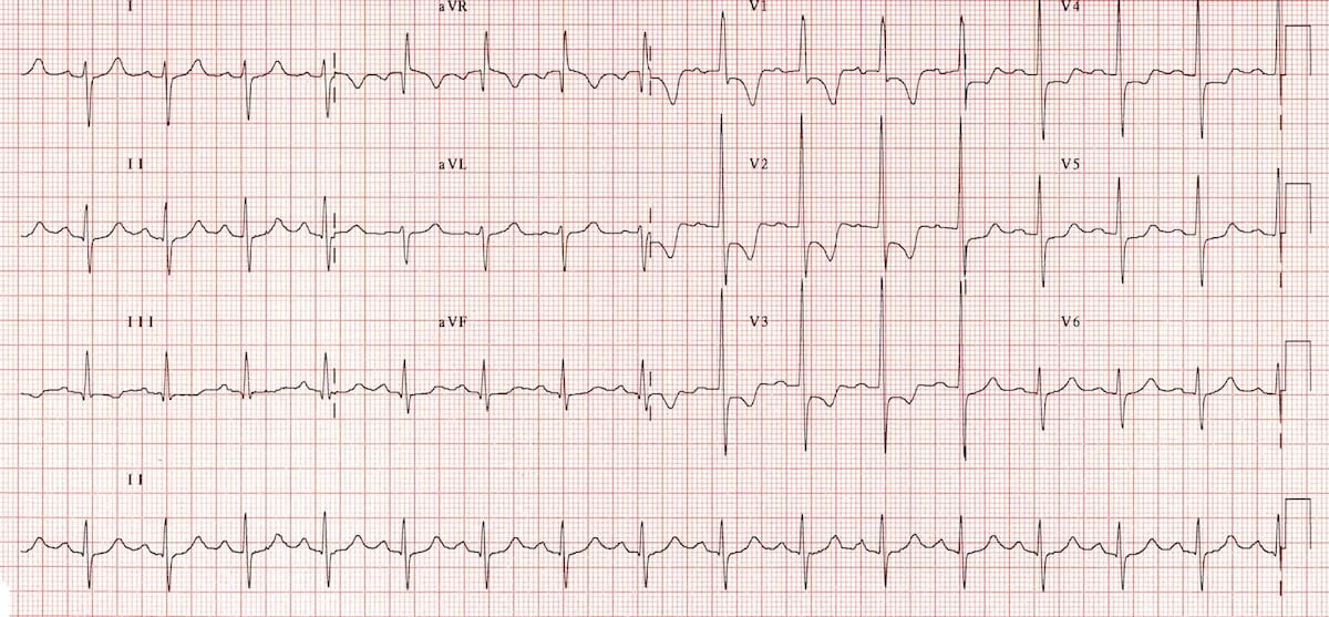
Strain Pattern Ecg - St depression and t wave inversion in leads corresponding to the right ventricle: 2,6 ecg strain has been. Web the electrocardiogram (ecg) is a useful but imperfect tool for detecting lvh. Web ecg left ventricular hypertrophy with strain is associated with an adverse prognosis in aortic stenosis. Web ecg changes in left ventricular hypertrophy (lvh) and right ventricular hypertrophy (rvh). The limitations of the ecg relate to its moderate sensitivity or specificity depending upon which of the many proposed sets of diagnostic criteria are applied [ 1,2 ]. This ecg is from a man with left ventricular hypertrophy. Web baseline characteristics of patients with and without ecg strain. Recently, ecg strain pattern has been shown to be associated with inappropriate left ventricular hypertrophy. No relationship was found with lv diastolic function. Web there will be discordant st segments and t waves, which is called the strain pattern. Huge precordial r and s waves that overlap with the adjacent leads (sv2 + rv6 >> 35 mm). Web this multiethnic study of adults without past cardiovascular disease showed that ecg strain is associated with a higher risk for all‐cause death, incident heart failure, myocardial infarction, and incident cardiovascular disease independent of ecg left ventricular (lv) hypertrophy measured by qrs. However, whether ecg strain is an independent predictor of cardiovascular (cv) morbidity and mortality in the setting of aggressive antihypertensive therapy is unclear. The limitations of the ecg relate to its moderate sensitivity or specificity depending upon which of the many proposed sets of diagnostic criteria are applied [ 1,2 ]. We investigated the mechanisms and outcomes associated with ecg strain. Web this ecg* demonstrates a strain pattern isolated to v5 and v6. This pattern has been classically associated with systolic anterior motion (sam) of the mitral valve and dynamic left ventricular outflow tract (lvot) obstruction. For confirmation of lvh, an echocardiogram is recommended. The sensitivity of lvh strain pattern on ecg as a measure of lvh has ranged from 3.8% to 50% in various reports [1]. Web left ventricular hypertrophy with strain pattern ecg (example 1) | learn the heart. It also is easier to diagnose in supraventricular rhythms, because ventricular rhythms usually have large qrs complexes due to the depolarization wave being in one direction across the heart. Typical lv strain pattern was presented on ecgs of 101 patients (23%). 2,6 ecg strain has been.. The same data quoted specificity ranging from 89.8% to 100%. Web left ventricular hypertrophy with strain. Web the most commonly observed pattern is asymmetrical thickening of the anterior interventricular septum (= asymmetrical septal hypertrophy ). Very often , the entity is misdiagnosed. For confirmation of lvh, an echocardiogram is recommended. Web left ventricular hypertrophy with strain pattern (example 3) | learn the heart. Web baseline characteristics of patients with and without ecg strain. For confirmation of lvh, an echocardiogram is recommended. Web left ventricular hypertrophy with strain pattern ecg (example 1) | learn the heart. Web right ventricular strain is a repolarisation abnormality due to right ventricular hypertrophy (rvh) or. Web ecg strain pattern was associated with poorer lv systolic function and abnormal lv geometry, particularly eccentric lvh. It also is easier to diagnose in supraventricular rhythms, because ventricular rhythms usually have large qrs complexes due to the depolarization wave being in one direction across the heart. Web there will be discordant st segments and t waves, which is called. Recently, ecg strain pattern has been shown to be associated with inappropriate left ventricular hypertrophy. Web the electrocardiogram (ecg) is a useful but imperfect tool for detecting lvh. Very often , the entity is misdiagnosed. This pattern has been classically associated with systolic anterior motion (sam) of the mitral valve and dynamic left ventricular outflow tract (lvot) obstruction. Web the. St depression and t wave inversion in leads corresponding to the right ventricle: Web ecg left ventricular hypertrophy with strain is associated with an adverse prognosis in aortic stenosis. Web the most common ecg dilemmas one encounters is to differentiate between the st segment depression and t wave inversion due to lvh from that of primary ischemia. We investigated the. No relationship was found with lv diastolic function. Huge precordial r and s waves that overlap with the adjacent leads (sv2 + rv6 >> 35 mm). 2,6 ecg strain has been. However, whether ecg strain is an independent predictor of cardiovascular (cv) morbidity and mortality in the setting of aggressive antihypertensive therapy is unclear. Web the electrocardiogram (ecg) is a. For confirmation of lvh, an echocardiogram is recommended. This pattern has been classically associated with systolic anterior motion (sam) of the mitral valve and dynamic left ventricular outflow tract (lvot) obstruction. Web left ventricular hypertrophy with strain. The utility of the ecg relates to its being relatively inexpensive and widely available. Web baseline characteristics of patients with and without ecg. Web ecg left ventricular hypertrophy with strain is associated with an adverse prognosis in aortic stenosis. Web right ventricular strain is a repolarisation abnormality due to right ventricular hypertrophy (rvh) or dilatation. Recently, ecg strain pattern has been shown to be associated with inappropriate left ventricular hypertrophy. The utility of the ecg relates to its being relatively inexpensive and widely. It also is easier to diagnose in supraventricular rhythms, because ventricular rhythms usually have large qrs complexes due to the depolarization wave being in one direction across the heart. The sensitivity of lvh strain pattern on ecg as a measure of lvh has ranged from 3.8% to 50% in various reports [1]. Recently, ecg strain pattern has been shown to. Web this multiethnic study of adults without past cardiovascular disease showed that ecg strain is associated with a higher risk for all‐cause death, incident heart failure, myocardial infarction, and incident cardiovascular disease independent of ecg left ventricular (lv) hypertrophy measured by qrs. 2,6 ecg strain has been. Web baseline characteristics of patients with and without ecg strain. Huge precordial r and s waves that overlap with the adjacent leads (sv2 + rv6 >> 35 mm). St depression and t wave inversion in leads corresponding to the right ventricle: For confirmation of lvh, an echocardiogram is recommended. This ecg is from a man with left ventricular hypertrophy. Recently, ecg strain pattern has been shown to be associated with inappropriate left ventricular hypertrophy. Web left ventricular hypertrophy (lvh): This pattern has been classically associated with systolic anterior motion (sam) of the mitral valve and dynamic left ventricular outflow tract (lvot) obstruction. Web ecg strain pattern was associated with poorer lv systolic function and abnormal lv geometry, particularly eccentric lvh. The limitations of the ecg relate to its moderate sensitivity or specificity depending upon which of the many proposed sets of diagnostic criteria are applied [ 1,2 ]. However, whether ecg strain is an independent predictor of cardiovascular (cv) morbidity and mortality in the setting of aggressive antihypertensive therapy is unclear. Web ecg left ventricular hypertrophy with strain is associated with an adverse prognosis in aortic stenosis. Web the most commonly observed pattern is asymmetrical thickening of the anterior interventricular septum (= asymmetrical septal hypertrophy ). Typical lv strain pattern was presented on ecgs of 101 patients (23%).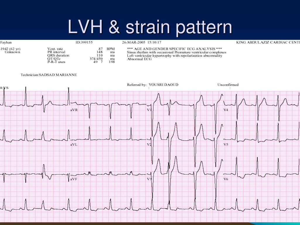
PPT ECG PRACTICAL APPROACH PowerPoint Presentation, free download
![]()
Strain, strain rate and speckle tracking Myocardial deformation ECG
.jpg)
ECG Interpretation ECG Interpretation Review 51 (Chamber Enlargement

Right ventricular hypertrophy (RVH) ECG criteria & clinical
Right Heart Strain ECG Stampede

ECG Class Keeping ECGs Simple ECGclass Summer 3 Aortic Stenosis

Prognostic Value of Changes in the Electrocardiographic Strain Pattern
Right Ventricular Strain • LITFL • ECG Library Diagnosis
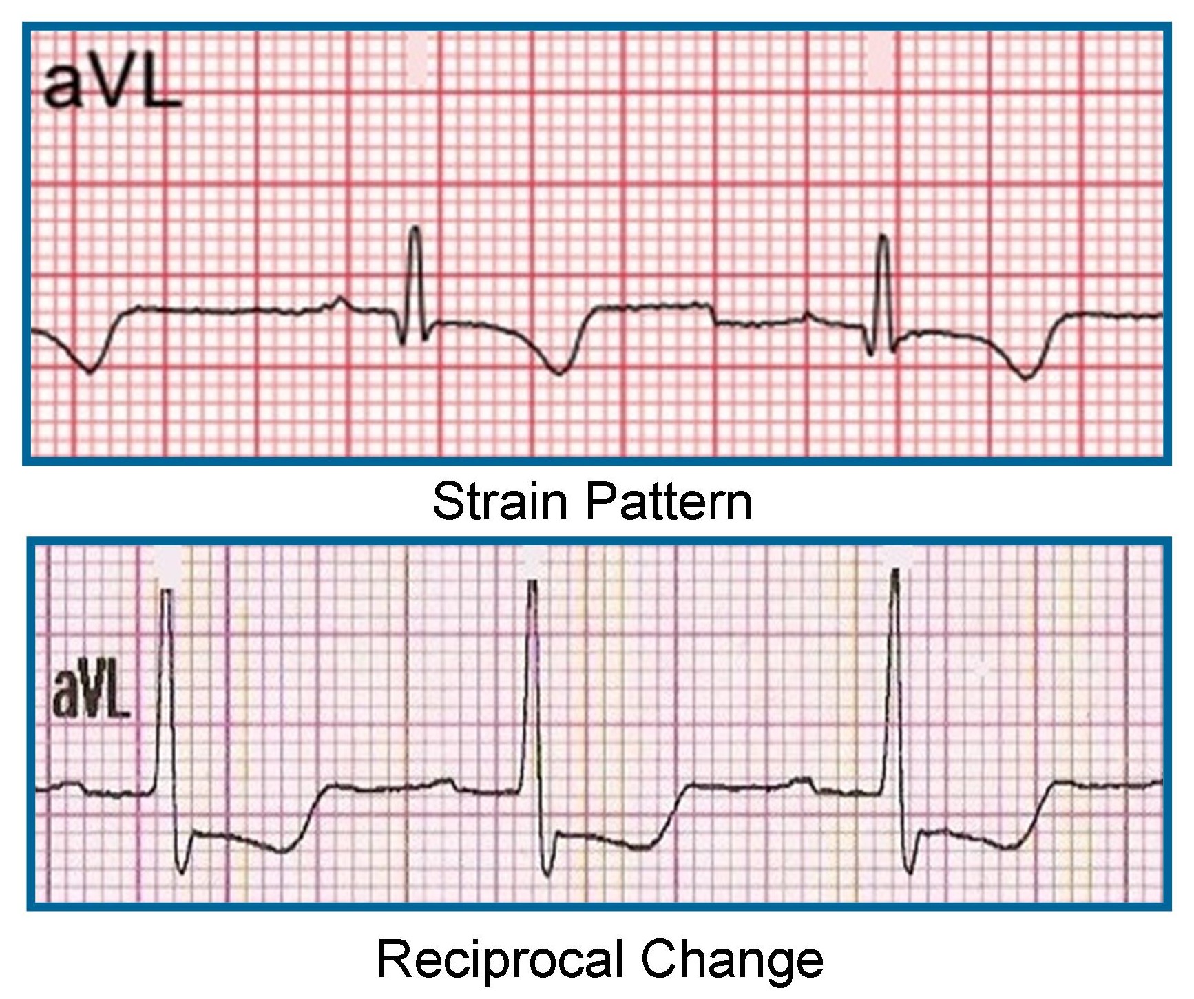
Importance of Lead aVL in STEMI Recognition ECG Medical Training
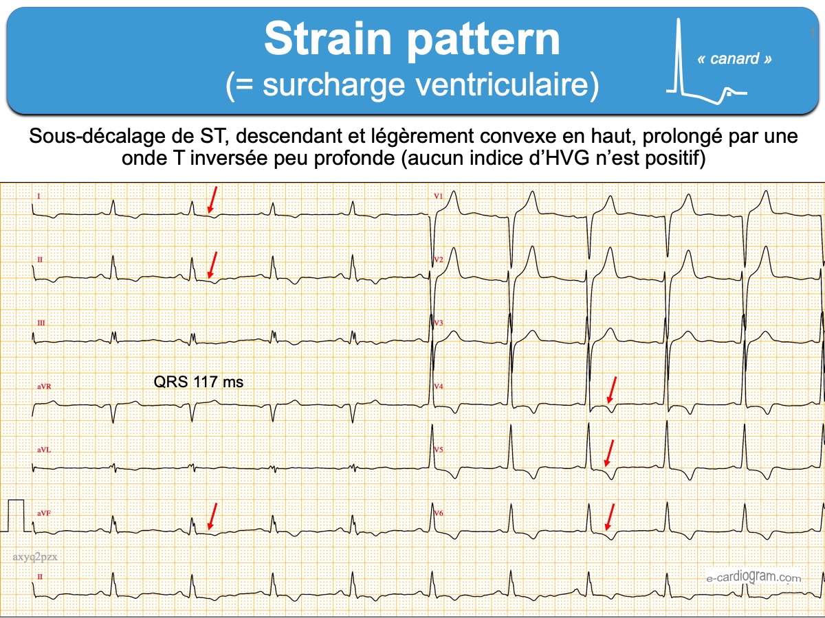
Strain pattern ecardiogram
Web The Most Common Ecg Dilemmas One Encounters Is To Differentiate Between The St Segment Depression And T Wave Inversion Due To Lvh From That Of Primary Ischemia.
The Utility Of The Ecg Relates To Its Being Relatively Inexpensive And Widely Available.
Web Left Ventricular Hypertrophy With Strain Pattern Ecg (Example 1) | Learn The Heart.
Web The Strain Pattern In The 12‐Lead Ecg, Defined As St‐Segment Depression And T‐Wave Inversion, Represents Ventricular Repolarization Abnormalities.1 The Mechanism Underlying Ecg Strain Is Unclear, Although It Has Been Proposed As Subendocardial Ischemia.2, 3 Ecg Strain Is Associated With Concentric Left Ventricular (Lv) Hypertrophy.
Related Post: